10. Critical Care for Trauma
Educational materials and pathways regarding the evaluation and management of the critically ill.
- Adult ICU Electrolyte Replacement
- Evaluation and Management of Atrial Fibrillation
- Nebraska Medicine Brain Death Criteria
- Percutaneous Tracheostomy Protocol
Adult ICU Electrolyte Replacement
Purpose
To define patients eligible for the electrolyte replacement protocol; to define the process for a provider to order the electrolyte replacement protocol; for a nurse to order and administer electrolyte replacement using this protocol; for a pharmacist to ensure safe dosing of electrolyte replacement; and for when the provider should be contacted when a patient has the electrolyte replacement protocol order set placed.
Policy
Standardized electrolyte replacement will be available for eligible adult ICU patient using an interdisciplinary approach. This includes but is not limited to medication management and monitoring.
Exclusion criteria are as follows:
- Pediatric patients (less than 19 yrs of age)
- Weight < 40 kg
- Renal dysfunction (serum creatinine 1.5 mg/dL or greater or increase in serum creatinine by 50% or renal replacement therapy) within the past 3 days
- pH <7.2 or pH >7.5 within the past 24 hours
- Diabetic ketoacidosis
Procedure
- The ICU Electrolyte Replacement Order Set will be initiated by the ordering provider. The provider will select which electrolytes (magnesium, potassium) they would like to have replaced via protocol, as well as the goal electrolyte level and preferred route of replacement.
-
- NOTE: if exclusion criteria has been met, the provider will be unable to place the order.
-
- The ICU Electrolyte Replacement Order Set will be continued perpetuity and should be evaluated daily to ensure appropriateness of continuation. If a patient develops exclusion criteria and the electrolyte protocol is still ordered, the nurse will be notified of the exclusion criteria that the patient has met and will be instructed to contact the provider regarding replacement.
- With the provider initiating and signing the ICU Electrolyte Replacement order, this allows the nurse to enter appropriate replacement and laboratory monitoring orders.
- When entering subsequent orders the nurse will enter those orders using the appropriate provider name and "Per protocol: cosign required".
Magnesium Replacement
-
-
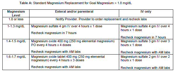
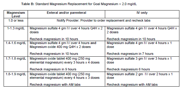
- The ICU Magnesium Replacement Order Set will be initiated by the ordering provider. They will be required to select the preferred route of replacement (enteral/parenteral or IV only) as well as the magnesium goal level.
- With the provider initiating and signing the ICU Electrolyte Replacement Order Set, this allows the nurse to enter the appropriate replacement and laboratory monitoring orders per Table A or B.
- when entering subsequent orders, the nurse will enter those orders using the appropriate provider name and "Per protocol: cosign required".
- If the Magnesium Replacement Order Set is initiated and the patient has sub-therapeutic magnesium levels within the previous 3 hours, a task will be added to the nursing work list.
- To address the magnesium electrolyte replacement, the nurse will access the ICU Electrolyte Replacement Order Sets within the manage orders tab. The order set will be listed under suggestions. Upon opening the order set, appropriate replacement and lab orders will be presented to the nurse per Table A or B, to enter and sign.
- During verification, the pharmacist will confirm that the order is appropriate per Table A or B.
-
- Duplicate replacement orders will flag on the verification screen.
- "Off protocol" oral replacement will be allowed in certain instances (i.e., continuation of home scheduled magnesium regimen).
-
- After pharmacist verification and acknowledgement of the order, the nurse will administer the ordered dose orally or via the infusion pump.
- The following situations describe when the ordering provider or designee MUST be contracted:
-
- The patient meets exclusion criteria and is ineligible to receive ongoing electrolyte replacement via this protocol.
- The magnesium level is below threshold specified by Table A or B.
-
-
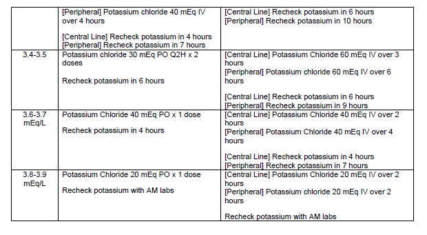
Potassium Replacement
-
-
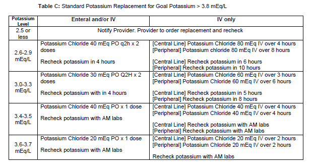
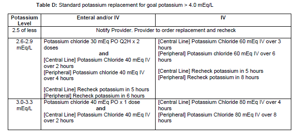
- The Potassium Replacement Order Set will be initiated by the ordering provider. They will be required to select the preferred route (enteral/parenteral or IV only) as well as the potassium goal level.
- With the provider initiated and signing the ICU Electrolyte Replacement Order Set, this allows the nurse to enter appropriate replacement and laboratory monitoring orders per Table C or D.
- When entering subsequent orders, the nurse will enter those orders using the appropriate provider name and "Per protocol: cosign required".
- If the Potassium Replacement Order Set is initiated and the patient has sub-therapeutic potassium levels within the previous 3 hours, a task will be added to the nursing work list.
- To address the potassium electrolyte replacement, the nurse will access the ICU Electrolyte Replacement Order Sets within the manage orders tab. The order set will be listed under suggestions. Upon opening the order set, appropriate replacement and lab orders will be presented to the nurse per Table C or D, to enter and sign.
-
- NOTE: if the RN has central line access, but is unable to administer using the central line due to concomitant infusions, they will contact the pharmacist to request a change in concentration.
- NOTE: if the RN has central line access, but is unable to administer using the central line due to concomitant infusions, they will contact the pharmacist to request a change in concentration.
-
- During verification the pharmacist will confirm that the order is appropriate per Table C or D.
-
- Duplicate replacement orders will flag on the verification screen.
- "Off protocol" oral replacement will be allowed in certain instances (i.e., continuation of home scheduled potassium regimen or intermittent loop diuretic doses).
-
- After pharmacist verification and acknowledgement of the order, the nurse will administer the ordered dose orally or via the infusion pump.
- The following situations describe when the ordering provider or designee MUST be contacted:
-
- The patient meets exclusion criteria and is ineligible to receive ongoing electrolyte replacement via this protocol.
- The potassium level is below threshold specified by Table C or D.
-
-
Authors
- Medication Management Committee (06/2022)
- P&T Formulary Committee (06/2022)
- Clinical Governance (07/2022)
Last Updated
7/2022
Evaluation and Management of Atrial Fibrillation
Purpose
- Establish a unified guideline for the diagnosis and treatment of new-onset atrial fibrillation (AF) in Acute Care Surgery patients.
Background/Definitions
- Primary AF: AF with no precipitating cause
- Secondary AF: AF precipitated by a secondary or reversible condition (e.g., surgery, sepsis, acute MI, etc. --most of our ICU patients)
Inclusion Criteria
- Patients with new onset atrial fibrillation.
Exclusion Criteria
- Patients with chronic atrial fibrillation.
Diagnostic Evaluation
- History:
- previous history of arrhythmia?
- currently on anticoagulation?
- Physical:
- irregular heart rhythm
- Imaging/Labs/Tests:
- ECG
- BMP+Mg+Phos
- Other labs at discretion of provider (CBC, blood cultures/infectious work-up, cardiac enzymes, etc)
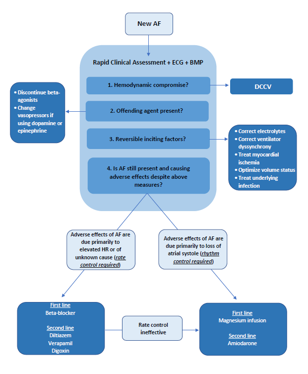
Practice Recommendations for Management
- New-onset, secondary AF is an organ dysfunction that signals something is wrong--need to address underlying cause while seeking to control rate/rhythm.
- Helpful questions to guide initial approach of patient with AF:
- 1) is the AF causing an immediate problem?
- 2)why is AF happening now (is this primary or secondary AF)?
- 3) should I worry about longer-term problems from the AF?
- Is the AF causing an immediate problem?
- When to consider rhythm control first:
-
- Emergent AF with severe decompensation:
-
- hypotension (SBP<100 or <110 for patients 65 and older), acute heart failure, altered mental status, cardiac ischemia
- if yes --> DCCV (direct current cardioversion)
- consider pairing DCCV with anti-arrhythmic such as amiodarone to increase probability of longer-term success.
-
- Non-emergent AF:
-
- consider a rhythm control strategy first if you think the patient needs atrial kick (i.e. severe mitral stenosis, aortic stenosis) or cannot tolerate nodal blocker (Wolf Parkinson White Syndrome)
-
- Emergent AF with severe decompensation:
-
- When to consider rate control first:
-
- Note: in most instances you can use rate control FIRST.
- Heart rate is higher than it would be with acute illness, but not immediately life threatening to require DCCV.
- Patient has contraindications to anticoagulation.
- Evidence to support a rate control strategy first during secondary AF: success of DCCV is low in secondary AF (as in ICU) --43% at 1 hr, 23% at 24 hrs remain in NSR.
-
- When to consider rhythm control first:
- Why is AF happening now?
- Fix electrolytes (magnesium is an effective rhythm control treatment).
- Fix volume status.
- Look for untreated infection.
- Remove beta-agonists.
- Should I worry about long-term problems from the AF?
- Arterial thromboembolism and AF recurrence are long-term concerns after new-onset AF in critically ill patients
- 44% af AF recurrence in 1 year after new-onset AF in sepsis.
- Cardiology follow-up (either inpatient or outpatient) for long-term rhythm monitoring and treatment plan should be considered.
Outcome Measures and Guideline Adherence
- AF (arrhythmia) is a PI filters for Trauma and Critical Care Surgery that is actively tracked/monitored.
Related Policies
Key Contributors
- Keely Buesing ,MD, FACS, Acute Care Surgery Division
Last Updated
February, 2023
References
- 2019 AHA/ACC/HRS Update
- 2014 AHA/ACC/HRS Guideline
- Um K et al. Pre- and post-treatment with amiodarone for elective electrical cardioversion of atrial fibrillation: a systematic review and meta-analysis. Europace. 2019;21(6):856-863.
- Arrigo M et al. Disappointing success of electrical cardioversion for new-onset atrial fibrillation in cardiosurgical ICU patients. Crit Care Med. 2015;43(11):2354-2359.
- Walkey AJ et al. Practice patterns and outcomes of treatments for atrial fibrillation during sepsis: a propensity-matched cohort study. Chest. 2016;149:74-83.
- Bosch NA et al. Comparative effectiveness of heart rate control medications for the treatment of sepsis-associated atrial fibrillation. Chest. 2021;159(4):1452-1459.
- Davey MJ et al. A randomized controlled trial of magnesium sulfate, in addition to usual care, for rate control in atrial fibrillation. Ann Emerg Med. 2005;45(4):347-353.
- Onalan O et al. Meta-analysis of magnesium therapy for the acute management of rapid atrial fibrillation. Am J Cardiol. 2007;99(12):1726-1732.
- Bosch NA et al. Atrial fibrillation in the ICU. Chest. 2018;154:1424-1434.
Supplemental Materials
- “Etiology of Atrial Fibrillation” schematic.
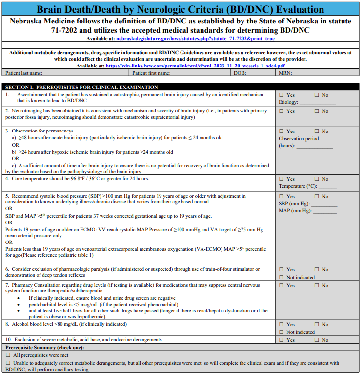
Nebraska Medicine Brain Death Criteria
Nebraska Medicine Policy Number: MS 29
Purpose
To give an accurate and complete description required to establish a diagnosis of breath death/Death by Neurological Criteria (BD/DNC), and to describe the roles and responsibilities of various clinicians and staff members in the process.
Scope
This policy applies to all patients at least 37 weeks corrected gestational age or older at Nebraska Medicine for whom a diagnosis of BD/DNC is considered.
Background
Nebraska Medicine follows the definition of BD/DNC as established by the State of Nebraska in statute 71-7202 and utilizes the accepted medical standards for determining BD/DNC.
A diagnosis of breath death is a clinical diagnosis that can only be established by a staff physician with privileges in neurology or critical/intensive care medicine. The staff physician will document the results of the brain death evaluation in the medical record. The time of death is determined at the time the evaluation is complete. Physicians in training, who are at an advanced level of training and deemed appropriate by the staff physician and working under the staff physician's direct supervision, can perform parts of the examination. The staff physician is fully responsible for the diagnosis, declaration, and documentation of brain death.
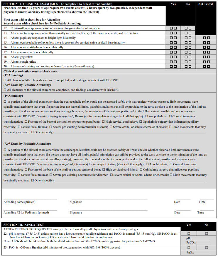
Brain Death Evaluation
A complete brain death evaluation consists of three components. All three components must be completed to establish a diagnosis of brain death:
- Establish permanent and proximate cause of coma
- Establish absence of cortical function and brain stem reflexes by neurologic examination
- Establish absence of spontaneous respirations by performing an apnea test
Completion of the three components of the brain death evaluation is sufficient to establish a diagnosis of brain death.
Ancillary Testing
Ancillary testing is not required if all three of the above components are completed. Ancillary tests may be used to support the diagnosis of brain death when uncertainty exists about the reliability of parts of the neurologic exam, when parts of the exam cannot be performed, or to shorten the interval between exams. the current acceptable ancillary tests are: Cerebral angiography, cerebral scintigraphy, and transcranial doppler (if age appropriate).
The interpretation of these tests must be interpreted by a staff physician with the required level of expertise.
Special circumstances:
- Physicians with recognized or potential conflicts of interest in relation to the outcome of the patient's care must remove themselves from the BD/DNC evaluation. For instance, a transplant service physician whose patient expires and has the potential for organ donation should excuse himself/herself from declaring the patient brain dead.
References
- Nebraska State Statute 71-7202. Determination of death. Source: Laws 1992, LB 906, 2.
- Pediatric and Adult Brain Death/Death by Neurologic Criteria Consensus Guideline. Neurology. Dec 12, 2023 issue: 101(24):1112-1132. Greer DM, Kirschen MP, Lewis A, Gronseth GS, Rae-Grant A, Ashwal S, Babu MA, Bauer DF, Billinghurst L, Corey A, Partap S, Rubin MA, Shutter L, Takahashi C, Tasker RC, Varelas PN, Wijdicks E, Bennett A, Wessels SR, Halperin JJ.
- The 2023 AAN/AAP/CNS/SCCM Pediatric and Adult Brain Death/Death by Neurologic Criteria Consensus Practice Guideline. A Comparison with the 2010 and 2011 Guidelines. Ariane Lewis, MD https://orcid.org/0000-0002-075807320, Matthew P. Kirschen MD, PhD https://orcid.org/0000-0003-358502687, and David Greer, MD https://orcid.org/0000-0002-2026-8333 AUTHORS INFO & AFFILIATIONS. December 2023 issue.
Related Policies and Procedures
Acute Bereavement Care -- TX02
Staff Accountability:
- Critical Care Medicine (09/2024)
- Medical Ethics Committee (09/2024)
- Pediatric Quality Committee (11/2024)
- Medical Staff Bylaws Committee NMC (11/2024)
- Medical Staff Medical Executive Committee NMC (11/2024)
- Board of Directors (11/2024)
Percutaneous Tracheostomy Protocol
Purpose
To provide guidance on safe practices to perform percutaneous tracheostomy in the operating room and ICU settings.
Preprocedural Planning
- Indications for tracheostomy at the discretion of the attending surgical intensivist.
- The procedure should be scheduled through the operating room by calling OR Charge Nurse.
- Two Attending Physicians must present at bedside to safely perform the procedure. One provider with perform the tracheostomy and one will be managing sedation. A procedure note and a sedation note need to be completed upon completion of the tracheostomy.
- Bronchoscopic guidance is required
- Medications for the procedure consist of an anxiolytic, a narcotic pain medication, and a neuromuscular paralytic. Additionally, local anesthetic may be requested.
- An intubation/airway cart with associated equipment is required at the bedside should reintubation or emergent airway be needed.
Equipment
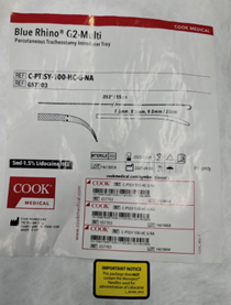
- Blue Rhino Percutaneous Tracheostomy Kit
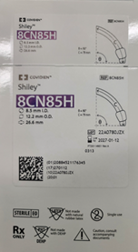
- Cuffed Tracheostomy (Size 6 and/or Size 8)
- Sterile Drapes and Chlorohexidine Prep
- Sterile Gowns and Gloves
- Eye protection and Head Coverings
- Bronchoscope
- Sterile water and Lubrication
- Airway cart (associated supplies needed for reintubation if needed)
Team
- Attending Intensivist to perform the tracheostomy
- Attending Intensivist to manage sedation
- Trainees (ICU Fellows, Surgical Residents, Medical Students)
- Critical care Nurse
- Respiratory Therapist
- OR Nursing Staff
Room Set-up and Patient Positioning
- The patient should be positioned supine with the neck slightly hyperextended with a shoulder roll if possible. If there is concern for or confirmed cervical spine injury, inline stabilization with the neck in a neutral position must be maintained with securely placed tape.

- The patient’s arms should be placed at patient’s side to ensure access to the neck bilaterally. The bed needs to be positioned to allow for access to the head of the bed so that orotracheal reintubation can be performed if needed.
- For right-handed surgeons, the bronchoscopy cart is generally placed on the patient’s left with the person performing the tracheostomy on the patient’s right. The respiratory therapist should be at the head of the bed with easy access to the patient’s airway. A second provider will be at the head of the bed performing the bronchoscopy. The patient’s nurse needs to have easy access to the patients IV in order to administer medications in a timely manner and the monitor with vital signs and pulse oximetry with audio easily visible to all.

- Set the ventilator to deliver a set volume and rate with 100% FiO2 to preoxygenate the patient. The ICU monitor should set so the pulse oximeter is audible. Continuous hemodynamic monitoring should be achieved with ekg and arterial line or frequent BP cuff monitoring (every 3 minutes).
Technical Steps
- Prior to the start of the procedure, a through “timeout” should be performed. All members of the team should be present and attentive.
- Adequate sedation should be achieved with anxiolytic and narcotic pain medications. This is followed by paralysis.
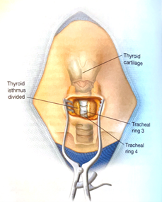
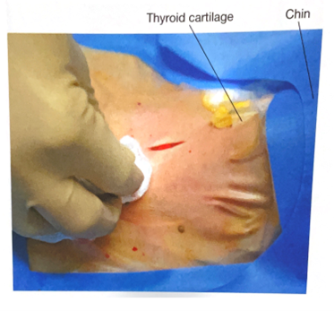
- Palpate the neck to identify relevant anatomy. Ideal location for placement of the tracheostomy is between the 2nd and 3rd tracheal ring.

- Don all appropriate PPE. Standard sterile surgical technique should be implemented.
- Surgically prepare neck and upper chest with chlorohexidine skin prep. Standard sterile technique and draping should be performed. Consideration for easy access to the endotracheal tube to allow for easy airway exchange after trach is placed.
- Anesthetize the skin and subcutaneous tissue with local anesthetic.

- Using a #15 scalpel, make a 2-3 cm vertical, midline incision approximately 40 mm cephalad (1-2 finger breaths) to the sternal notch and just below the cricoid cartilage. If an anterior jugular vein is encountered in the incision (even if no injury is suspected), consider ligation proximally and distally as this is easiest to perform before the tracheostomy tube has been placed.

- Using a hemostat, bluntly dissect the subcutaneous tissue and muscle in midline to the pretracheal tissue along the length of the incision to better palpate the trachea to determine the point of entry.
- With the bronchoscope adaptor in place, advance the bronchoscope into the airway. Inspect the trachea and bronchial trees and clear any secretions.
- With the assistance of the respiratory therapist, while keeping the bronchoscope at the end of the endotracheal tube, retract both the endotracheal tube and bronchoscope simultaneously until the subglottic structures are visualized and one can see the anterior wall of trachea being palpated by the surgeon. The bronchoscope should be always kept within the endotracheal tube during this portion of the procedure in order to maintain control of the airway and ensure that the bronchoscope is not damaged.
- ***Although usually unnecessary, cautery may be used prior to entering the trachea with the introducer needle. After entry into the trachea, cautery should not be used do to the risk of fire with open oxygen source.***
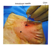
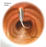
- An introducer needle is used the enter the anterior portion of the trachea between the 2nd and 3rd tracheal ring (approximately 1 finger breadth below the cricoid cartilage). With the bevel of the needle facing downward, the guidewire is passed into the trachea. Visualization of the guide wire going in the direction of the carina is required. Advance the guidewire slightly passed the carina into the right or left mainstem bronchus.


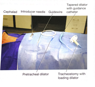
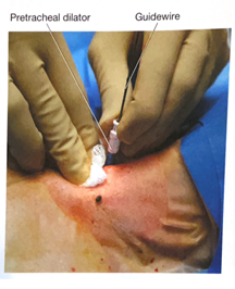
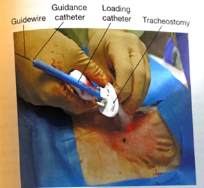
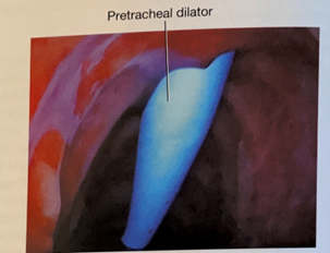
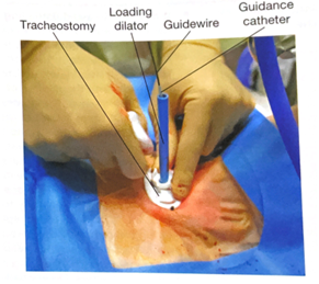
- Using the Seldinger technique, with constant Bronchoscopic visualization and control of the wire within the trachea, the trachea is sequentially dilated. The dilator handle is hydrophobic which makes it less likely to slip in a wet environment while the actual dilating portion is hydrophilic which only requires water/liquid to be lubricated. First the small tracheal dilator is advanced over the wire to dilate the pretracheal tract. Next the single-stage tapered dilator and the guiding catheter are advanced as a unit over the wire to dilate the trachea. Markings on the side of the progressive dilators guide the depth to which they are inserted. All catheters (pretracheal dilator, tapered dilator, and guiding catheter) should enter perpendicular to the trachea as to prevent pretracheal dissection or false passage. If the patient has limited ventilatory reserve prior to the procedure, the bronchoscope can be removed prior to the dilation portion of the procedure.


- The tapered dilator is removed from the guiding catheter and the guidewire, leaving the guiding catheter and the guidewire in place. If there is a longer distance between the tracheal surface and the skin surface, a finger can be used to dilatate the tract to help facilitate placement of the tracheostomy during the next step.
- Next, an appropriately sized and well lubricated tracheostomy tube with introducer is advanced over the wire and guiding catheter into the trachea. The wire, guiding catheter and loading trocar is then removed, keeping the tracheostomy in place.


- Inflate the tracheostomy cuff, insert the inner canula and connect tracheostomy to ventilator circuit. The presence of end-tidal carbon dioxide after ventilation resumes confirms placement in the airway.
- A bronchoscopy should be performed through the newly placed tracheostomy to visually confirm that it is within the trachea in proper position. Only remove ET tube after placement of tracheostomy tube within the trachea is confirmed.
- The tracheostomy is secured with a tracheostomy collar or ties to help prevent accidental dislodgement and provide time for adequate to tract formation.
- Obtain a chest x-ray to confirm appropriate positioning of the tracheostomy tube, rule out pneumothorax, and evaluate for bronchial obstruction.


Tracheostomy Care
- Following tracheostomy placement, standardized tracheostomy care bundles should be implemented. These protocols include steps to ensure the tracheostomy is secure, suctioning techniques, daily stoma hygiene and knowledge of emergency protocols should the newly placed airway be compromised. Special attention should be given to prevent pressure ulceration, particularly along the inferior aspect of the tracheostomy faceplate, especially if the flange is sutured to the skin.
- The tracheostomy tube should be exchanged no sooner than post-operative day 7.
- Once the patient is liberated from the ventilator and secretions are reasonably managed the process of tracheostomy tube downsizing can occur. The downsizing if the tracheostomy tube allows for improved patient comfort and the ability to participate in speech therapy.
- After the tracheostomy is no longer necessary, the patient can be decannulated. The stoma is covered with sterile occlusive dressing generally closes within two to four days.
Key Contributors
Bennett Berning, MD
Last Updated
March, 2023
References
- Cheung NH, Napolitano LM. Tracheostomy: epidemiology, indications, timing, technique, and outcomes. Respir Care. 2014 Jun;59(6):895-915; discussion 916-9.
- Young D, Harrison DA, Cuthbertson BH, Rowan K, TracMan Col- laborators. Effect of early vs late tracheostomy placement on survival in patients receiving mechanical ventilation: the TracMan randomized trial. JAMA 2013;309(20):2121-2129.
- Holevar M, Dunham JC, Brautigan R, Clancy TV, Como JJ, Ebert JB, Griffen MM, Hoff WS, Kurek SJ Jr, Talbert SM, Tisherman SA. Practice management guidelines for timing of tracheostomy: the EAST Practice Management Guidelines Work Group. J Trauma. 2009 Oct;67(4):870-4.
- Delaney A, Bagshaw SM, Nalos M. Percutaneous dilatational tracheostomy versus surgical tracheostomy in critically ill patients: a systematic review and meta-analysis. Crit Care 2006;10(2):R55.
- Hashimoto DA, Axtell AL, Auchincloss HG. Percutaneous Tracheostomy. N Engl J Med. 2020 Nov 12;383(20):e112.
- Hawn, M. T., Berning, B. J., & de Moya, M. A. (2023). Tracheostomy: Open and Percutaneous. In Operative techniques in surgery (2nd Edition, Vol. Volume Two, pp. 2506–2512). Wolters Kluwer.